Figure 3.
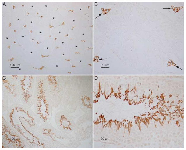
RHBG and RHCG localization in the testis. (A and B) Low- and high-power micrographs respectively of RHBG immunolabeling in the testis. No RHBG immunolabel is present in seminiferous tubules (asterisks). However, strong plasma membrane RHBG immunolabel is present in interstitial cells with morphology and localization consistent with identification as Leydig cells (arrows).
No immunolabel was observed in experiments in which primary antibody was omitted (data not shown). (C and D) Representative low- and high-power micrographs respectively of RHCG immunolabel in the testis. RHCG exhibits significant variation in immunolabel intensity in different seminiferous tubules, consistent with stage-dependent variations in RHCG expression. High-power micrographs (D) spermatids, but not Leydig cells, display RHCG immunolabel. Controls in which primary antibody was omitted had no detectable immunolabel. Results are representative of findings from at least four mice.

