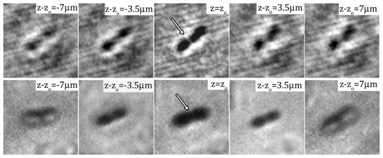
Fig. 5.

Numerically reconstructed images of a single E. coli cell recorded by DHM at the magnification of 40X with the range of at the interval of 3.5 . The center image, i.e. , is the in-focus image. The distance from the in-focus plane is marked on each sub image. The bottom row shows microscopic images of an E. coli at the corresponding planes from the in-focus location recorded by Nikon TiE with a Nikon Plan Fluor 40X objectives.
