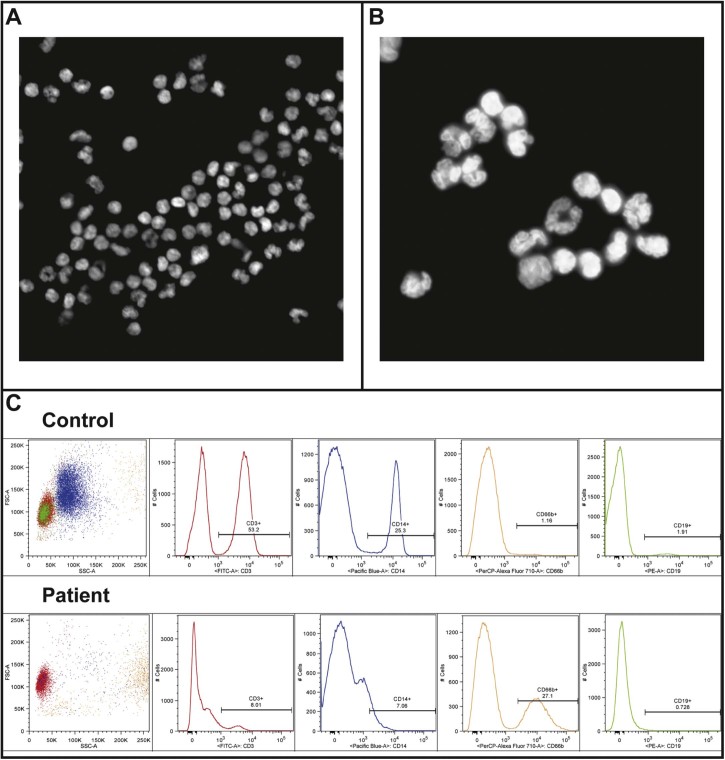
Fig. 5.
Blood cell distributions and nuclear lobulation in the patient. A) Dapi staining of nuclei from cells isolated from patient blood, 20× magnification. B) Dapi staining of nuclei from cells isolated from patient blood, 100× magnification. C) FACS staining of cells isolated from patient and control blood for following markers: CD3 (T-cells), CD14 (macrophages), CD66b (granulocytes) and CD19 (B-cells).

