Figure 2.

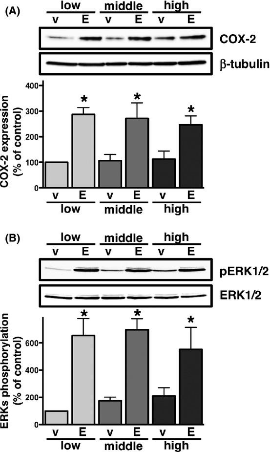
Effects of cellular density on EGF-induced COX-2 expression (A) or phosphorylation of ERKs (B) in HCA-7 cells. Cells were cultured at low (3 × 10⁴ cells/each 6 well), middle (1 × 10⁵ cells/each 6 well), and high (3 × 10⁵ cells/each 6 well) densities. Each cellular density was treated with either vehicle (v) or 50 ng/mL of EGF (E) for 6 h (A) or 15 min (B) at 37°C and then subjected to immunoblot analysis with an antibody against COX-2 (A) or phospho-ERKs 1 and 2 (pERK1/2) (B) as described in the Materials and Methods. The blots were stripped and re-probed with an antibody against β-tubulin (A) or ERKs 1 and 2 (ERK1/2) (B). The bar graphs represent the ratio of COX-2 to β-tubulin (A) or pERK1/2 to total ERK1/2 (B) as assessed with pooled densitometric data (mean ± SD) from more than three independent experiments. Data are normalized to the ratio of COX-2 to β-tubulin (B) or pERK1/2 to total ERK1/2 (C) of vehicle-treated controls at the low cellular density as 100%. *P < 0.05, ANOVA, significantly different from the vehicle treatment.
