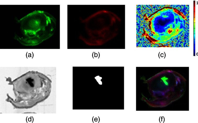
Fig. 5.
A single cross-sectional slice of the entire mouse head is shown with brain region in the upper mid-half. (a) Characteristic untargeted tracer slice image. (b) Characteristic targeted tracer slice image. (c) Binding potential image resulting from snapshot processing of (a) and (b). (d) Green fluorescent protein highlighting image. (e) Segmentation of tumor site from GFP image. (f) RGB overlay with the targeted image in the red channel, the GFP image in the green channel, and the untargeted image in the blue channel.

