
Figure 2.
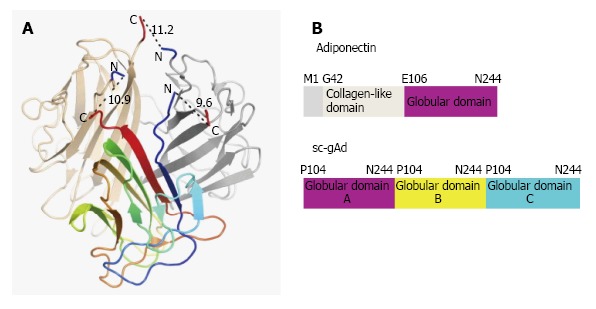
Structure of single-chain globular domain adiponectin (sc-gAd). A: Base region of mouse gAd structure where blue arrow determines the N terminus and red arrow determines the C terminus; B: Domain organization of human adiponectin and the sc-gAd, where there are three domains A, B and C respectively. (Adapted from Min et al[30]).