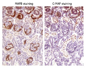
Figure 2.

Immunostaining for MAFB and c-MAF in fetal human kidney tissue. An immunohistochemical analysis was performed using primary antibodies against MAFA (BL1069; Bethyl Laboratories, Inc.), MAFB (P20; Santa Cruz Biotechnology, Inc.), and MAF c-Maf (M153; Santa Cruz Biotechnology, Inc.). The details are described in reference[40]. A sample of human normal fetal kidney tissue (male, 25 wk) was purchased from BioChain (catalog No. T8244431, Lot No. A606275). Glomerular podocyte lesions stained positive for MAFB, and while the proximal tubules stained positive for c-MAF.
