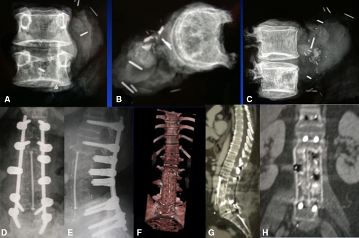
Fig. 2A–H.
Postoperative images are shown for the patient in Fig. 1. (A–C) CT scan shows the tumor immediately after en bloc removal to assess margins and histopathology, which reported wide margins. (D, E) Postoperative radiographs show the reconstruction of the defect. (F) Three-dimensional, (E) sagittal and (H) coronal CT scans taken at 18 months show no cage subsidence and good fusion. The patient was alive with no evidence of disease at 8 years.

