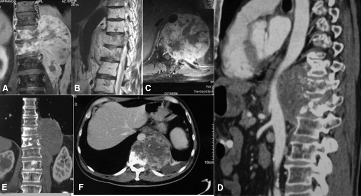
Fig. 3A–F.
Images illustrate the case of a 64-year-old man diagnosed with four-level (T10–L1) chordoma, with involvement of the adventitia of the aorta. (A) Coronal, (B) sagittal, and (C) axial MR images show a tumor spanning from T10 to L1. (D) A CT angiogram shows the tumor in close proximity to the aorta, involving its adventitia. (E) Coronal and (F) axial CT scans show the extent of the tumor. We undertook resection using an anterior–posterior approach. The adventitia of the aorta was adherent to the tumor. It was separated and the tumor was removed en bloc along with the adventitia via the posterior approach.

