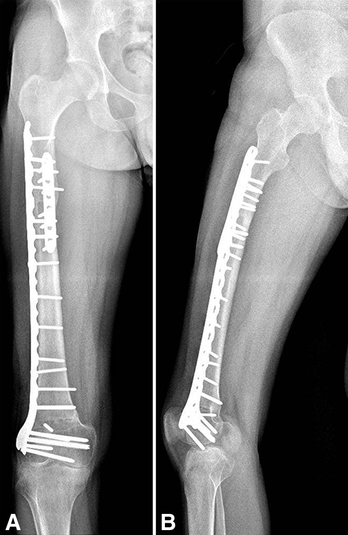
Fig. 2A–B.

AP and lateral 2-year postoperative radiographs are shown of the right femur after resection of the fractured allograft and reconstruction with a second intercalary allograft. (A) AP radiograph of the right femur showing healing of both osteotomies; a distal femur locking plate was used in the lateral side that covers both osteotomies and the addition of a short anterior plate in the proximal osteotomy. (B) Lateral radiograph of the right femur showing adequate fixation of both osteotomies.
