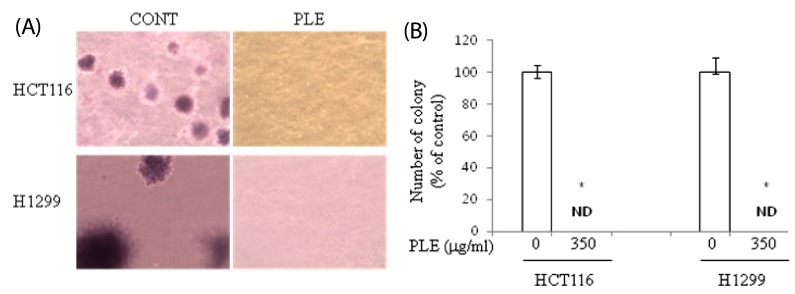
Fig. 2.
Effect of PLE on colony formation of human colon and lung cancer cells in soft agar. HCT116 and H1299 cells on agar were treated with PLE at the concentrations of 0 or 350 µg/ml for 21 days (A). Colonies were stained with crystal violet, and the representative area is shown (B). The number of colonies was counted in four randomly selected points in each well under phase contrast time-lapse microscopy (×100). Asterisks indicate statistical differences between untreated control and PLE-treated cells by two-tailed student t-test (P < 0.05). ND: not detected.

