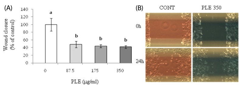
Fig. 5.
Effect of PLE on migration in human lung cancer cells. H1299 cells were treated with 0, 87.5, 175, and 350 µg/ml of PLE for 24 h. (A) The width of wound was quantified using Image-J software, and the wound closure during the 24 h time interval is shown as mean ± SE of 4-5 determinations. Different letters (a-c) indicate statistical differences among different concentrations by Tukey's test (P < 0.05). (B) Representative wound area of H1299 cells before and after 24 h treatment with PLE at the concentration of 350 µg/ml is shown.

