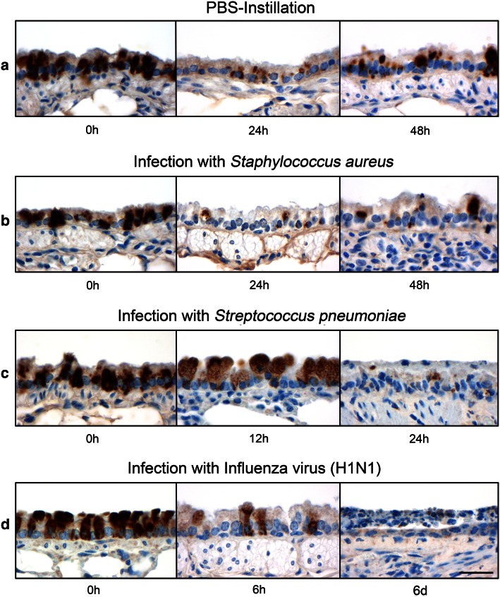
Fig. 4.
mCLCA5 protein expression disappeared in various challenge models. Lungs from naive (n = 4) and PBS-treated (n = 4) mice as well as from mice infected with S. aureus (n = 4), S. pneumoniae (n = 2) and influenza virus (n = 2) were examined at the extrapulmonary to intrapulmonary junction to characterize the presence and the course of mCLCA5 protein expression in this specific location at various time points. a, b Comparison of naive lungs to lungs from PBS-treated or S. aureus-infected mice revealed a significant reduction in mCLCA5 protein expression 24 h after infection, with a slight tendency toward a return after 48 h. c, d After infection with S. pneumoniae and influenza virus, the immunosignal of mCLCA5 disappeared over time. Bar 20 µm

