
Figure 6.
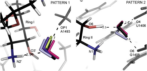
Patterns 1 (left) and 2 (right). Superposition of the A-site complexes showing water molecules mediating the interactions of RNA (gray) with ribostamycin (RIO (pink)), neomycin (NMY (violet)), paromomycin (PAR (black)), lividomycin (LIV (light gray)), modified paromomycin (JS4 (blue)), and tobramycin (TOY (light green)). PAR atoms are shown in black (carbon atoms), red (oxygen), blue (nitrogen), and white (hydrogen). The distances indicated by the dashed lines correspond to the PAR complex. To see this figure in color, go online.