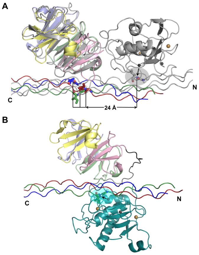
Figure 6. Collagen triple-helix positioning across MT1-MMP HPX domain and fulllength MMP-1.
(A) The complex of THP with HPX domain of MT1-MMP (colored as in Figure 5) is superimposed via the HPX domain with the NMR model of the THP complex with full-length MMP-1 (Bertini et al., 2012) colored gray. The scissile Gly-Ile peptide bond in the MMP-1 complex is shown with sticks within the gray oval. The horizontal line marks potential distances of translation between the scissile bond positions in the MT1-MMP complex and the MMP-1 complex.
(B) Illustration of the hypothesis that the catalytic domain (cyan) of MT1-MMP folds over the collagen triple-helix to form a sandwich with the HPX domain. The scissile bond is highlighted by an arrow and a cyan oval at the active site. Calcium and zinc ions are indicated by gold and gray spheres, respectively. Hypothetical paths of the interdomain linker are colored gray. See Movie S1 for a simulated, speculative trajectory of reorientation between the hypothetical modes of binding of panels A and B.

