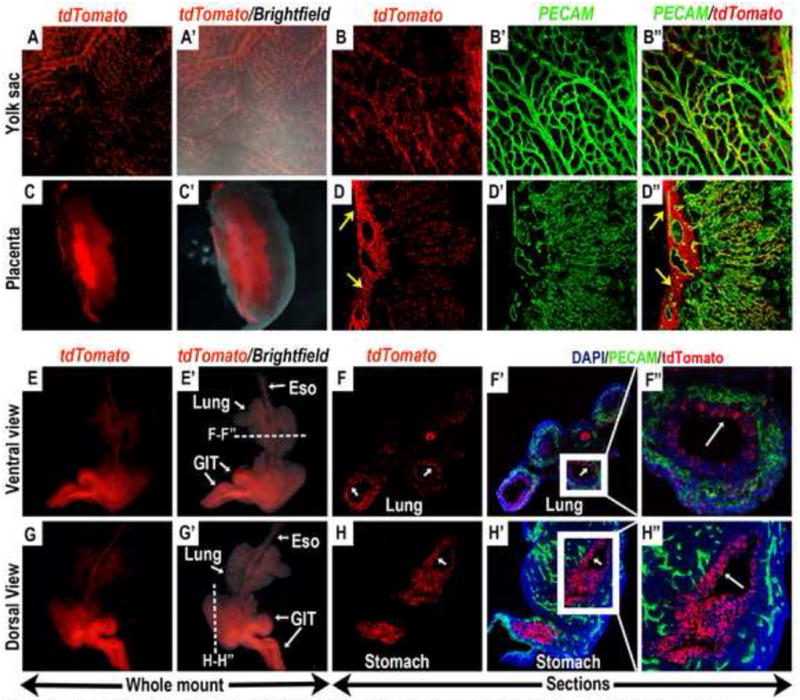
Figure 4. Contribution of E6.5 Wnt11-CreER expressing cells to the endothelium and endodermally derived internal organs in E12.5 embryos.

Wnt11-CreER mediated recombination and activation of tdTomato expression was induced by a single dose of tamoxifen administration at E6.5, and embryos were harvested and analyzed at E12.5. (A-D”) Whole mount epi-fluorescent analysis showed that tdTomato expressing cells were lining the yolk-sac vasculature (A and A’), and co-expressed the endothelial marker PECAM (B, B’ and B”). Bisection of the placenta revealed that widespread tdTomato expression could also be observed in the placental labyrinthe (C and C’). In the placenta, tdTomato expression was present mainly in the endothelial cells in the fetal vasculature (D’ and D”) as well as the supporting chorio-allanatoic mesenchyme (yellow arrows in D and D”) surrounding the larger placental vessels. (E-H”) In the embryo proper, tdTomato expressing cells contributed to the endodermally derived internal organs, namely the lungs and the gastro-intestinal tract. (E, E’, G, G’) whole mount epifluorescent analysis. Sectioning along the indicated planes in E’ and G’ (dashed white lines) revealed that the tdTomato expressing cells were present mostly in the epithelial lining of the lungs (white arrows in F and F’; magnified view in F”) and stomach (white arrow in H and H’; magnified view in H”). Eso: esophagus, GIT: gastro-intestinal tract.
