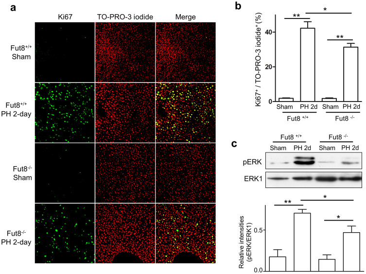
Figure 3. Cell proliferation was suppressed in the livers of Fut8−/− mice.
(a) Immunostaining for liver tissues (10 μm frozen section) of Fut8+/+ and Fut8−/− mice using anti-Ki67 antibody (200 × field). The positive cells of the immunostaining were labeled with the green spots (left panel), and the nuclei were labeled by TO-PRO-3 iodide (red spots, middle panel). (b) The quantitative data were obtained from at least 3 mice in each group. **, P < 0.01. (c) Equal protein of liver lysates at day 2 after PH were separated by 10% SDS-PAGE and blotted with anti-phospho-ERK and anti-ERK1 antibodies. The quantitative data were obtained from 3 mice in each group. *, P < 0.05, **, P < 0.01.

