
Figure 1. Tissue distribution of CIC and ATXN1L proteins in mice.
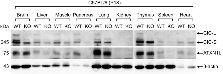
Western blot analysis for tissue distribution of CIC and ATXN1L proteins at P18. Twenty μg of total protein extract was loaded on each lane. KO means Cic-L-/- mouse.
Official websites use .gov
A
.gov website belongs to an official
government organization in the United States.
Secure .gov websites use HTTPS
A lock (
) or https:// means you've safely
connected to the .gov website. Share sensitive
information only on official, secure websites.
Western blot analysis for tissue distribution of CIC and ATXN1L proteins at P18. Twenty μg of total protein extract was loaded on each lane. KO means Cic-L-/- mouse.