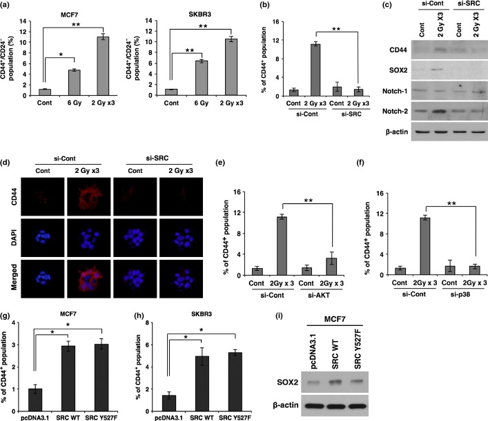
Figure 4.
Irradiation promotes breast cancer stem cell populations through SRC signaling. (a) Quantification of CD44+/CD24− cell population by FACS analysis in MCF7 and SKBR3 cancer cells after irradiation. (b) Quantification of CD44+ cell population by FACS analysis in MCF7 cancer cells transfected with siRNA targeting SRC (si-SRC) or scrambled control siRNA (si-Con) prior to irradiation. (c, d) Western blot analysis for CD44, SOX2, Notch-1, and Notch-2 (c), and immunocytochemistry for CD44 (d) in MCF7 cancer cells transfected with siRNA targeting SRC or scrambled control siRNA prior to irradiation. (e, f) Quantification of the CD44+ cell population by FACS analysis in MCF7 cancer cells transfected with siRNA targeting AKT (si-AKT) (e) or p38 MAPK (si-p38) (f) prior to irradiation. (g, h) Quantification of the CD44+ cell population by FACS analysis in MCF7 (g) and SKBR3 (h) cancer cells 48 h after transfection with SRC WT, mutant form SRC Y527F, or control vector pcDNA3.1. (i) Western blot analysis for SOX2 in MCF7 cells 48 h after transfection with SRC WT, mutant form SRC Y527F, or control vector pcDNA3.1. β-actin was used as a loading control. Error bars represent mean ± SD of triplicate samples. *P < 0.05; **P < 0.01. Cont, control.

