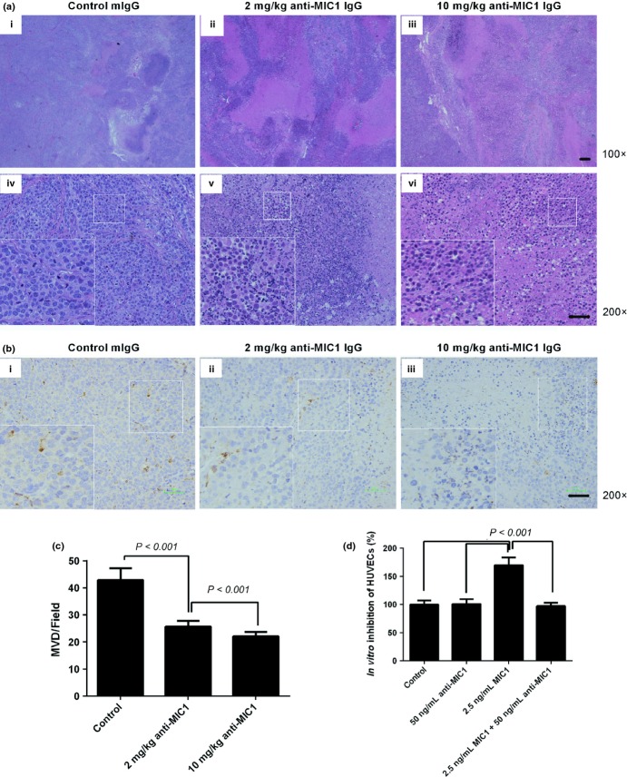
Fig. 5.
The tumor tissue necrosis and inhibition of angiogenesis in vivo. (a) Representative hematoxylin and eosin-stained tumor tissues obtained from mice in the control (i, iv), 2 mg/kg treated group (ii, v) and 10 mg/kg treated group (iii, vi). (b) Detection of blood vessels via immunohistochemical staining for Von Willebrand factor (VWF) from the control group (i), 2 mg/kg treated group (ii), 10 mg/kg treated group (iii). (c) Quantification of angiogenesis assessed by microvessel density (MVD). Data represent the mean ± standard error (SE), n = 4. (d) The effect of macrophage inhibitory factor (MIC) and anti-MIC1 antibody on human umbilical vein endothelial cells (HUVECs) in vitro; Data represent the mean ± SE, n = 4.

