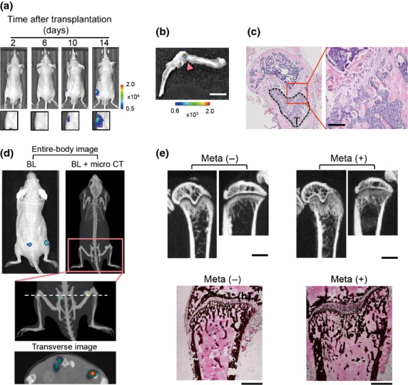
Figure 1.

Murine osteosarcoma LM8 cells develop osteoblastic bone metastasis. (a) Representative time course bioluminescence (BL) images after intracardiac (i.c.) transplantation of LM8/luc. (b) Ex vivo imaging of LM8/luc tumor-bearing hind limb shown in (a) (14 days after LM8/luc injection). LM8/luc metastasis signal is indicated by an arrowhead. Scale bar = 5 mm. (c) Hematoxylin–eosin staining of hind limb bone with LM8 metastasis (T) of (b). Scale bar = 100 μm. (d) Multimodal imaging. Images were obtained 14 days after i.c. transplantation of LM8/luc. The dashed line indicates imaging section of the transverse image. Micro CT, micro X-ray computed tomography. (e) Aberrant bone formation due to osteoblastic bone metastasis in the femur and tibia. Micro X-ray CT images were obtained 21 days after i.c. injection of LM8 (upper panels). Scale bar = 1 mm. The lower panels indicate that von Kossa staining of the same metastasis-free (Meta−) and bone metastatic (Meta+) femurs as the upper panels. Scale bar = 500 μm.
