
Figure 4.
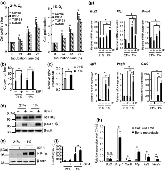
Insulin-like growth factor (IGF)/IGF receptor (IGFR) signaling and hypoxia cooperatively stimulate progression of murine osteosarcoma LM8 cells. (a) Proliferation assay of LM8/luc with IGF-1 (100 ng/mL), transforming growth factor-β1 (TGF-β1; 10 ng/mL), and receptor activator of factor-κB ligand (RANKL; 100 ng/mL) in 21% or 1% O2. (b) Colony formation assay of LM8/luc with IGF-1 (100 ng/mL) for 14 days in 21% or 1% O2. (c) Quantitative RT-PCR (qRT-PCR) analysis of Igf1r expression in LM8/luc cultured for 16 h in 21% and 1% O2. (d) Protein expression and phosphorylation levels of IGF1R. The cells were precultured for 16 h in 21% or 1% O2 then treated with IGF-1 (100 ng/mL) for 1 h. (e) Effect of IGF-1 on hypoxia-inducible factor-1α (HIF-1α) protein level. LM8 cells were treated with IGF-1 (100 ng/mL) for 12 h in 21% and 1% O2. (f) Effect of IGF-1 on HIF transcriptional activity. LM8/HRE-luc cells were treated with IGF-1 (100 ng/mL) for 12 h in 21% and 1% O2. *P < 0.05. (g) qRT-PCR analysis of downstream genes of IGF/IGFR signaling and HIF. LM8/luc cells were treated with IGF-1 (IF) (100 ng/mL) for 6 h in 21% and 1% O2. *P < 0.05. (h) qRT-PCR analysis of downstream genes of IGF/IGFR signaling and HIF in bone metastasis at 14 days after LM8 injection. *P < 0.05.