Figure 5.

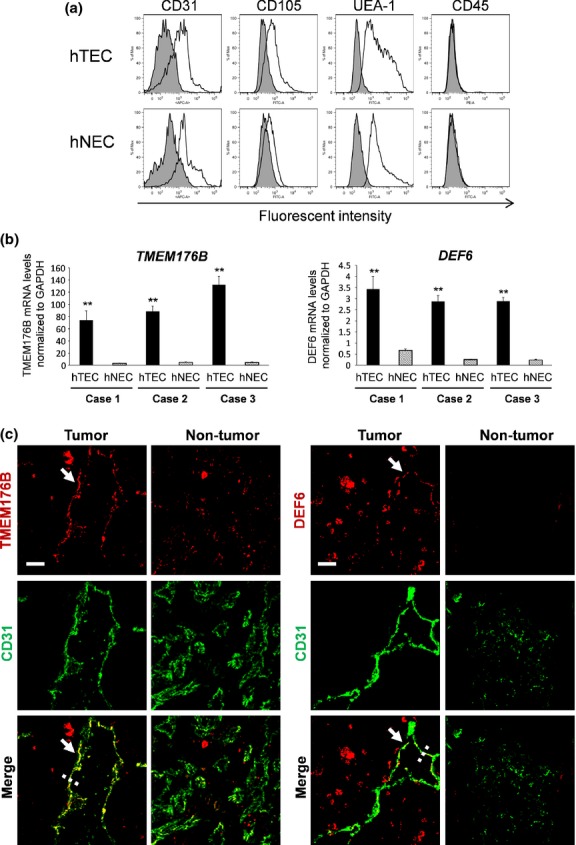
Analysis of TMEM176B and DEF6 expression in vitro and in vivo. (a) Verification of endothelial cells (EC) from a human sample. The binding of ulex europaeus agglutinin 1 (UEA-1 lectin), expression of CD31, CD105 and lack of expression of CD45 (white area) indicates high purity of the isolated human tumor endothelial cells (hTEC) and human normal endothelial cells (hNEC). The isotype control is shown in gray. (b) Upregulated expression of TMEM176B and DEF6 in hTEC. qRT-PCR analysis detected high levels of expression of both genes in hTEC compared with the corresponding hNEC in all three cases. Expression levels of the mRNA were normalized to that of GAPDH (**P < 0.01). (c) Both TMEM176B and DEF6 were strongly stained in tumor vessels using an anti-CD31 antibody in combination with an antibody against either TMEM176B or DEF6. In contrast, normal vessels (glomerular) of normal renal tissue were weakly stained. All samples were counterstained with DAPI. Profiles of immunofluorescence intensities along the dashed lines are shown in Figure S3(a,b). The signal intensities of TMEM176B or DEF6 in the CD31-positive area of whole sections were analyzed by ImageJ (NIH, Bethesda, MD, USA) quantitatively (Fig. S3c,d). Bar, 20 μm.
