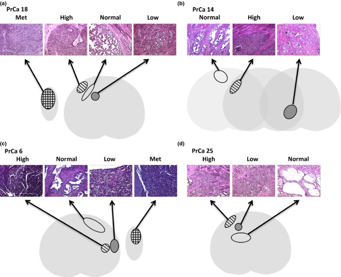
Fig. 1.
Histology of coincident prostate cancer foci. Representative H&E stains and illustrations representing the prostate cancer foci that were laser microdissected. Two cancer foci and uninvolved prostate glands were isolated from PrCa 18 (a), PrCa 14 (b), PrCa 6 (c) and PrCa 25 (d). In addition, a metastatic (Met) focus was isolated from PrCa 18 and PrCa 6. For PrCa 14 (b), the foci were from different levels of the prostate. For the other three specimens the foci were at the same level. Light gray, histologically normal prostate; dark gray, low-grade cancer focus; striped, high-grade cancer focus; checked, metastatic focus from a lymph node removed at the time of prostatectomy.

