Fig. 3.
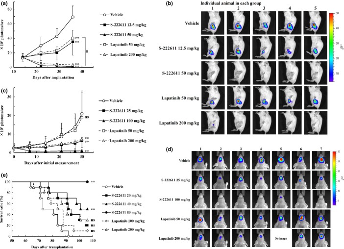
In vivo antitumor activity of S-222611 in patient-oriented preclinical models. (a–d) Luciferase-expressing human breast cancer cell line, BT474-luc, were implanted into the bone marrow of the left femur (a,b) or into the brain (c,d) of mice. (a,c) Tumor growth was monitored by the photons emitted from the tumor as an indicator, which was measured with an IVIS Imaging System 200 (Caliper Life Sciences). (b,d) Bioluminescent images at the last measurement. Tumor-bearing mice were orally administered vehicle or the indicated doses of S-222611 or lapatinib for 21 days (a,b) or 28 days (c,d). The photons were measured once or twice weekly and the mean photons/sec with SD are presented at each data point. a,b: n = 5, c,d: n = 7. In the experiment with intracranially implanted mice (c,d), 1 mouse treated with 200 mg/kg lapatinib died accidentally during the anesthesia for in vivo imaging at 13 days after the initial measurement. The death was not considered to be related to lapatinib treatment. Thus, the data points of the 200 mg/kg lapatinib group at 14 days after initial measurement represent the mean and SD of the data from 6 mice. ns: P ≥ 0.05, **: P < 0.01 (versus vehicle, Dunnett's test). #: P < 0.01 (50 mg/kg S-222611 vs 50 mg/kg lapatinib, Welch's t-test). (e) Human breast cancer cell line, BT474, were implanted into the brain of nude mice. After randomization at 36 days after implantation (n = 10), vehicle or multiple doses of S-222611 or lapatinib were orally administered daily until death or the end of study (106 days after implantation). ns: P ≥ 0.05, **: P < 0.01 (log-rank test with Dunn–Šidák multiple test correction).

