Fig. 4.
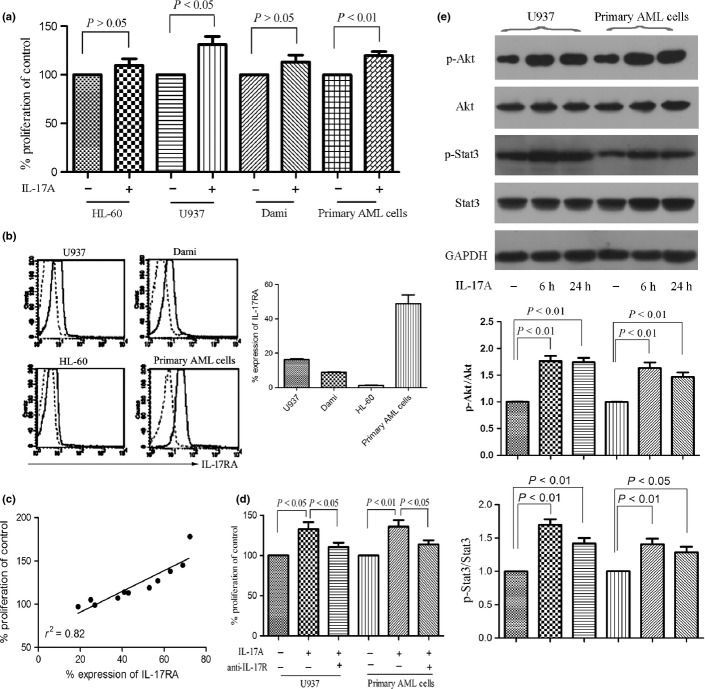
Interleukin (IL)-17A promotes the proliferation of AML cells via IL-17 receptor (IL-17R). (a) Acute myeloid leukemia (AML) cell lines HL-60, U937, Dami, and primary AML cells isolated from AML patients (n = 23) were incubated with or without IL-17A (50 ng/mL) for 7 days and proliferation was assayed by MTT. Data are showed in proliferation in the presence of IL-17A compared with control and expressed as mean ± SEM representing at least three independent experiments. (b) The IL-17RA expression of AML cells was measured using anti-CD217 PE antibody (solid line) or mouse IgG1 PE antibody (dotted line) by flow cytometry. Representative histograms (left panel) and statistical data (right panel) were shown. (c) A correlation was observed between the IL-17RA expression and the IL-17A-inducing proliferation. (d) The effects of IL-17R on U937 and primary AML cell proliferation were determined by incubating the cells with IL-17A (50 ng/mL) in the presence or absence of anti-IL-17R antibody (3 μg/mL) for 7 days. (e) Western blotting showed the phosphorylation of Akt and Stat3 were significantly increased after IL-17A stimulation for 6 h and lasted for 24 h in IL-17R+ AML cells. Representatives and statistical data were shown for four independent experiments.

