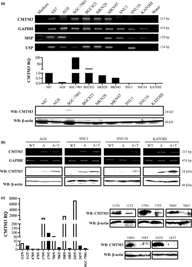
Figure 1.
(a) Expression and methylation status of CMTM3 in cell lines was detected by RT-PCR, methylation-specific PCR (MSP), unmethylation-specific PCR (USP), and quantitative PCR. Western blotting (WB) confirmed the expression of CMTM3. RQ, relative quantity. (b) Expression of CMTM3 was detected by RT-PCR and WB after treatment with demethylation agent. A, cells treated with 5-aza-2′-deoxycytidine; A+T, cells treated with 5-aza-2′-deoxycytidine and trichostatin A; WT, untreated cells; (c) CMTM3 expression in gastric cancer paired tissues was detected by quantitative PCR and WB. Normal mucosae tissues (N) are shown as white columns; tumor tissues (T) are shown as black columns. Gastric cancer cell lines were also analyzed with the same standard, and the relative quantity of SGC-7901 was present.

