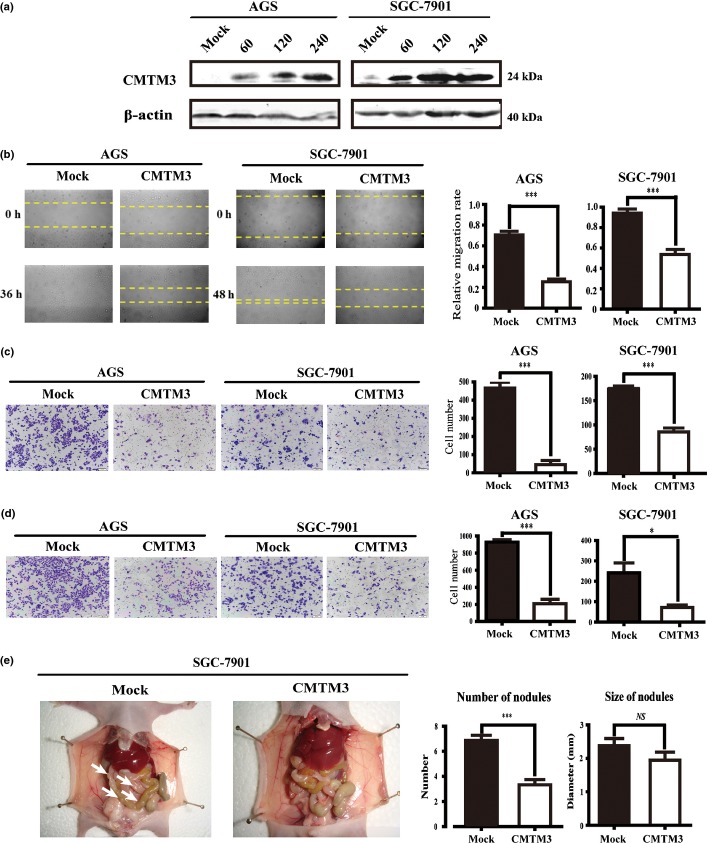
Figure 2.
(a) Overexpression of CMTM3 in SGC-7901 and AGS gastric cancer cells was detected by Western blot analysis. Different MOIs (0, 60, 120, and 240) of adenovirus were used. (b) Effect of CMTM3 on cell migration was observed by wound healing assay. Photographs were taken at indicated time points after the scratch (magnification, ×100). (c, d) Effects of CMTM3 on cell migration (c) and invasion (d) were detected by Transwell assay (magnification, ×100). The statistical graph indicates the mean ± SD and P-value of the number of cells per five random high power fields (magnification, ×400) counted from three independent experiments. (e) Ability of cell migration and invasion in vivo was detected by a peritoneal spreading model in nude mice. Photographs were taken after the mice were killed. Arrows indicate tumor nodules. NS, not significant. *P < 0.05; ***P < 0.001.

