
Figure 3.
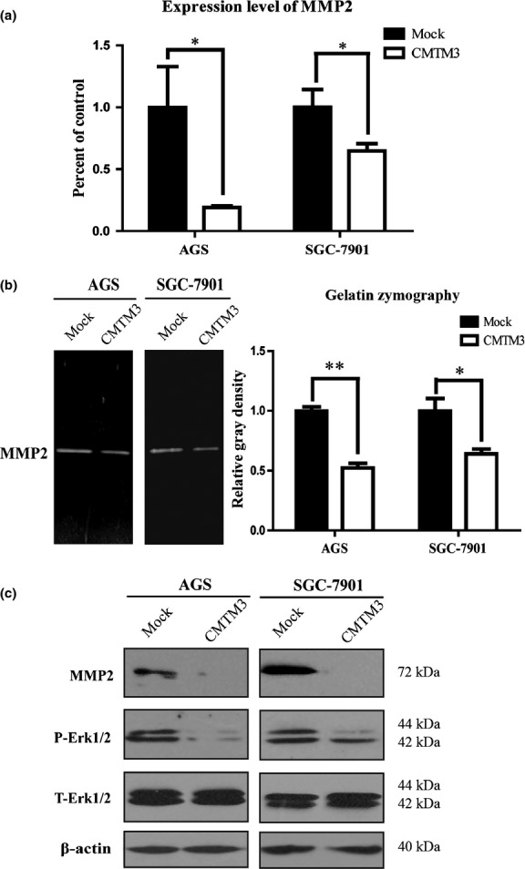
(a) Real-time PCR was carried out to detect MMP2 expression in AGS and SGC-7901 gastric cancer cells. Average percentage of control (Mock) with SEM is shown from three independent experiments. (b) MMP2 in AGS cell supernatants was observed by gelatin zymography. Average relative gray density with SEM is shown from three independent experiments. (c) MMP2, phosphorylated Erk1/2 (P-Erk1/2) and total Erk1/2 (T-Erk1/2) in two cell lines were observed by Western blotting. The experiment was repeated at least three times and one representative result is shown. *P < 0.05; **P < 0.01.