Figure 5.

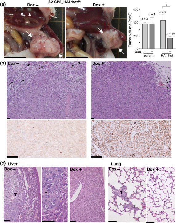
Intra-pancreatic transplantation of S2-CP8_HAI-1tet#1 cells in nude mice. The treated mice were maintained with or without Dox administration and killed 26 days after injection. (a) Macroscopic findings of pancreatic tumors (arrows). Metastases and infarction in the liver are indicated by arrow heads and asterisks, respectively. Bar, 1 cm. Primary tumor volumes of parent S2-CP8 and S2-CP8_HAI-1tet#1 cells without or with Dox treatment are also shown (mean ± SEM). *P < 0.001 (Mann–Whitney U-tests). (b) Histology of pancreatic tumors (upper panel, H&E) and immunohistochemistry of HAI-1 (lower panel). Arrows indicate pancreatic acinar tissue. Bars, 50 μm. (c) Histology of metastatic lesions (liver and lungs) formed in the absence of Dox treatment. T, metastatic tumor. Bars, 50 μm.
