
Figure 3.
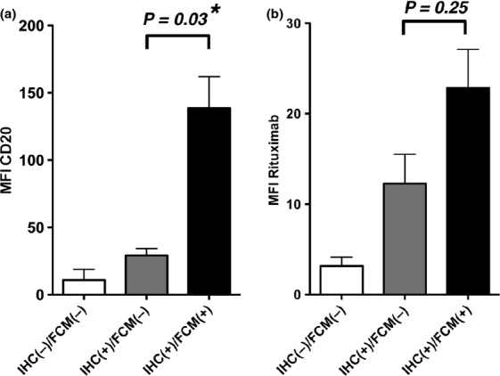
Flow cytometry (FCM) analyses using anti-CD20 B1 antibody and fluorescent-labeled rituximab. (a) FCM analysis using anti-CD20 B1 antibody was performed, and the MFIs of lymphoma cells were measured. RRBL1 and WILL2 cells were utilized as representative CD20 IHC(−)/FCM(−) samples. The P-value is shown, and the asterisk indicates a statistically significant difference. (b) The MFI value using Alexa 488-labeled rituximab was also analyzed in the same lymphoma samples as (a).