Figure 2.

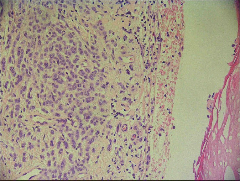
Skin biopsy showed an infiltrate of malignant cells characterized by pleomorphic atypical nuclei and ample pale cytoplasm (hematoxylin and eosin, ×2000)

Skin biopsy showed an infiltrate of malignant cells characterized by pleomorphic atypical nuclei and ample pale cytoplasm (hematoxylin and eosin, ×2000)