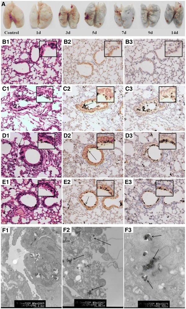
Figure 3.

Images of the lung tissue in mice and the IL-8 and IL-6 expression in the lung tissue of mice by immunohistochemical staining. A: Images of the lung tissue in mice after CB inhalation for 14 d. The mice were deeply anesthetized with chloral hydrate and perfused by injection from the left ventricle with 20 mL of 37°C saline solution and then the lung tissues were separated. B1-E1: Histopathology of the lung tissue in mice after exposure to CB particles for different time by HE staining (200×). The arrows in C2, D2, and E2 indicate the CB particles in pulmonary alveoli or bronchioli. The arrows in D1 and E1 indicated inflammatory cells. B2-E2: The IL-8 expression in lung cells after CB exposure for different time points by immunohistochemical staining (200×). The IL-8 positive cells displayed brownish yellow granules. In lung cells, IL-8 was located mainly in the cytoplasm and karyon. B3-E3: The IL-6 expression in lung tissue after CB exposure for different time points by immunohistochemical staining (200×). In lung cells, IL-6 was a granular brown substance located mainly in the cytoplasm and karyon. B1-B3: Control group; C1-C3: 7 d CB exposure group; D1-D3: 14 d CB exposure group; and E1-E3: recovery for 14 d after 14 d CB exposure. Inset: a higher magnification of the lung tissue (400×). F: TEM images of lung cells in mice after CB inhalation for 14 d. F1: Alveolar type II epithelial cells in control (5000×); F2: The disintegration of the macrophages in the lung of mice after CB exposure for 14 d (15000×) (Staining by uranyl acetate and lead citrate); and F3: The CB particles in a lung macrophage (25000×) (No uranyl acetate and lead citrate staining). The arrows indicate the CB particles in a lung macrophage.
