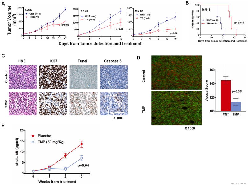
Figure 5. TMP inhibits MM cell growth and prolongs survival in vivo.

(A) U266, OPM2 ansd MM1s cells were injected s.c. in 3 different cohorts of SCID mice. Following detection of tumor, mice were treated with either 2 mg TMP or placebo s.c. daily for 3 weeks. Tumors were measured in two perpendicular dimensions once every 3 days. (B) Survival was evaluated from the first day of treatment until death using the Graphpad analysis software (C) Tumors were isolated from TMP-treated and control mice and sections were evaluated by histological examinations following Ki67 staining showing decreased proliferation; caspase3 and TUNEL stains showing significant tumor cell apoptosis. (D) Evaluation of survivin levels was performed in the paraffin embedded tumor sections stained with a combination of primary antibodies specific for CD138 and survivin. The tissue sections were imaged and the relative amount of survivin localized within tumor-cell nuclear compartment was determined using Automatated quantitation of antigen expression (AQUA) analysis. The data reflects the results of 12 fields imaged at 200x magnification per tumor. (E) In the SCID-hu model, mice, at the first detection of tumor, were treated with vehicle (n=3) or TMP (n=3). Serum samples were collected weekly and level of shuIL-6R was measured by ELISA as a marker of tumor growth. Baseline values before treatment were not significantly different among groups.
