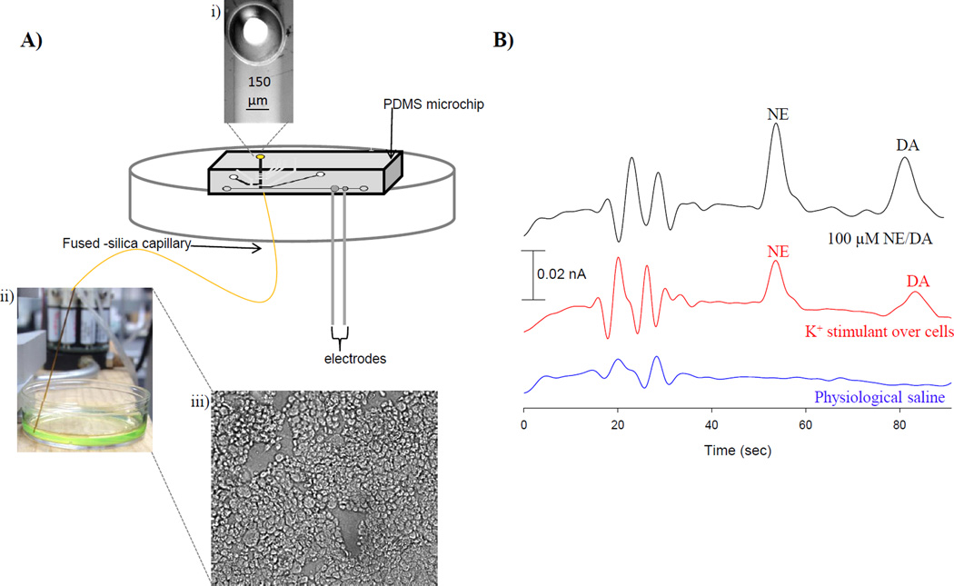
Figure 5.
PC 12 cell analysis. A) Schematic of assembly for PC 12 cell analysis. i) The PDMS microchip is sealed over the embedded fused-silica capillary. ii) The capillary is placed in solution and sample is withdrawn to the PDMS microchip. iii) Micrograph of immobilized PC 12 cells. B) Electropherograms of physiological saline, K+ stimulated release, and 100 µM NE/DA standards.

