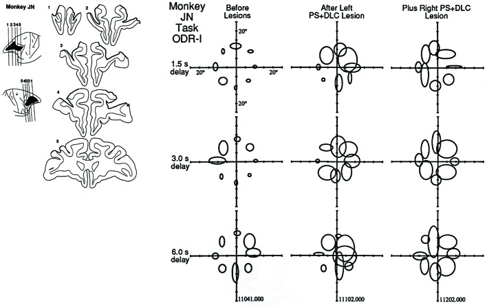
Figure 3.
Effects of focal lesions of the dorsolateral prefrontal cortex on performance in the ODR task. In this monkey (Monkey JN), the first lesion was located in the left principal sulcal region and, several months later, a second lesion was applied in the right principal sulcal region. The locations of the ellipses indicate the locations where the visual cues were presented. The size of the ellipse indicates the magnitude of the behavioral deficit. Three delay lengths (1.5 s, 3.0 s, and 6.0 s) were randomly applied. Note that larger deficits were observed when the visual cues were presented in the contralateral visual field with respect to the lesioned hemisphere. Reproduced from Funahashi et al. (1993a).

