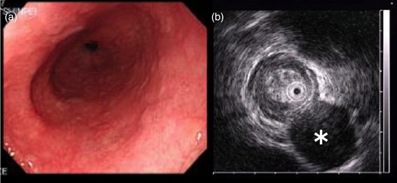
Figure 2:

Endoscopic examination and endoscopic ultrasonography. (a) Tumor with a normally appearing mucosa was located 40 cm from the incisor teeth. (b) Hypoechoic submucosal tumor (asterisk) with annular localization, arising from the submucosal layer.
