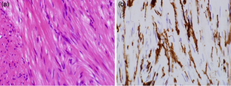
Figure 4:

Histological specimens prepared from the removed GIST. The left and right microphotographs were taken at a magnification of ×40. (a) Hematoxylin and eosin-stained specimen showed spindle cells. (b) Immunohistochemical-stained specimen showed that the cells were positive for c-KIT.
