Figure 6.
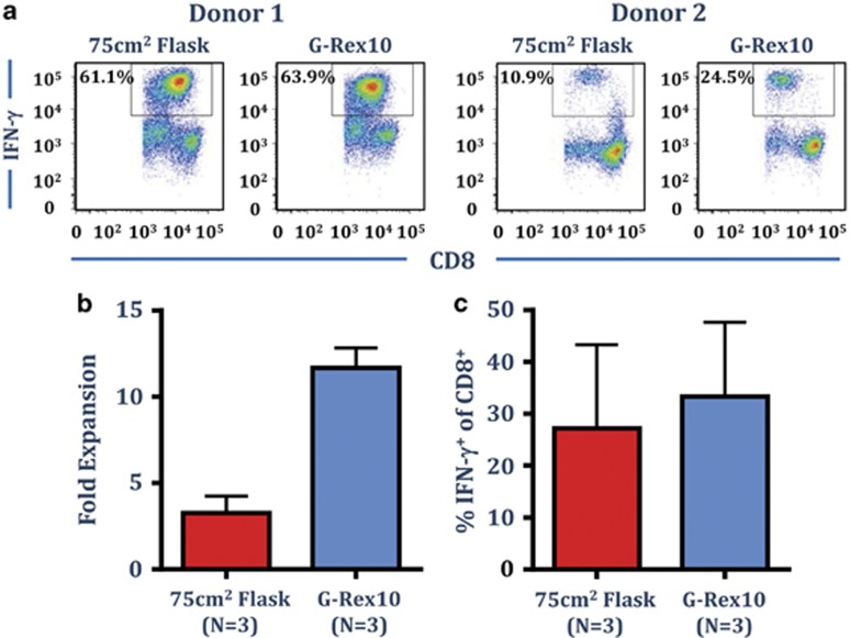
Optimal expansion of EBV LMP1&2- and EBNA1-specific T cells in OpTmizer supplemented with CTS Immune Cell SR in G-Rex10 flasks. PBMC from three EBV-seropositive donors were cultured with autologous AdE1-LMPpoly infected PBMC in OpTmizer-SR (final concentration 5%) in either a 75 cm2 flask or a G-Rex10 flask. T cell cultures were supplemented with 50% fresh media containing 120 IU ml−1 IL-2 after 3 days and every 3–4 days after. On day 14, cell numbers were determined using trypan blue exclusion, prior to determining T-cell specificity using an intracellular IFN-γ assay following recall with a pool of defined LMP1&2 and EBNA1 CD8+ T-cell peptide epitopes. (a) Representative intracellular IFN-γ analysis from two different donors is shown. (b) Data represent the mean±s.e.m. of the fold expansion of viable cells as determined by trypan blue. (c) Data represent the mean±s.e.m. of frequency of LMP1&2/EBNA1-specific CD8+ T cells.

