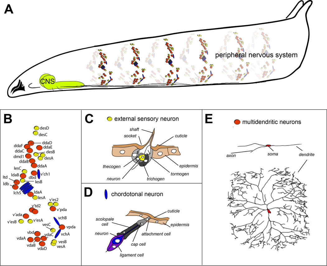
Figure 1. Organization of the embryonic and larval peripheral nervous system of Drosophila.
A. Drawing of a third instar Drosophila larva showing sensory elements that comprise the peripheral nervous system. For simplicity, only sensory neurons of a subset of abdominal segments are shown. Bundled sensory axons project to the central nervous system (CNS) that resides in the ventral and anterior part of the larva.
B. Schematic of the arrangement of sensory neurons in a single abdominal hemisegment. External sensory organs are indicated by yellow circles, chordotonal organs by blue ovals, and multidendritic neurons by red circles.
C. Drawing of external sensory organ structure. Names of individual cellular elements are indicated. Drawing adapted, with permission, from Comprehensive Molecular Insect Science. Vol. 1: Hartenstein V. Development of Insect Sensilla. pp. 379–419, 2005.
D. Drawing of chordotonal organ structure. Names of individual cellular elements are indicated. Drawing adapted, with permission, from Comprehensive Molecular Insect Science. Vol. 1: Hartenstein V. Development of Insect Sensilla. pp. 379–419, 2005.
E. Tracings of multidendritic neurons. Two different neurons are shown, the dorsal bipolar dendrite neuron (top), and a class IV nociceptive neuron (bottom). Note the different degrees of dendritic branching shown by the two neurons. Tracing of class IV neuron reproduced, with permission from Grueber et al., 2003.

