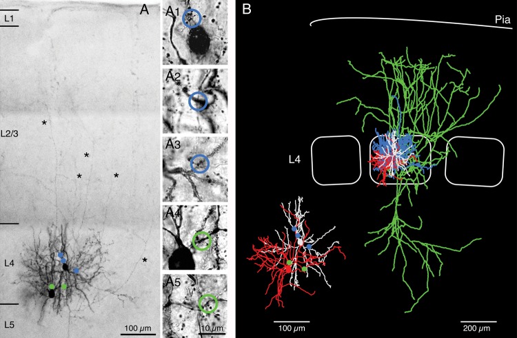
Figure 6.
Identification of synaptic contacts. (A) Low-power photomontage of a synaptically connected pair of a BIn and a spiny stellate cell that were filled with biocytin during recording. The 2 neurons showed reciprocal coupling. Putative, light microscopically identified GABAergic synaptic contacts between the presynaptic L4 interneuron (left) and the postsynaptic spiny stellate cell (right) are marked by green dots. Putative reciprocal excitatory synaptic contacts are indicated by blue dots. Asterisks highlight long axonal collaterals of the spiny stellate cell. (A1–5) High-power images of synaptic contacts. Blue open circles, inhibitory contacts (A1–3); green open circles, excitatory contacts (A4 and A5). (B) Neurolucida reconstruction of the same neuron pair shown in (A). Blue, L4 interneuron axon; red, L4 interneuron soma and dendrite; green, L4 spiny stellate axon; white, L4 spiny stellate soma and dendrite. The inset shows the dendritic arbor of pre- and postsynaptic neuron together with the putative synaptic contacts (same color code as for panel A).

