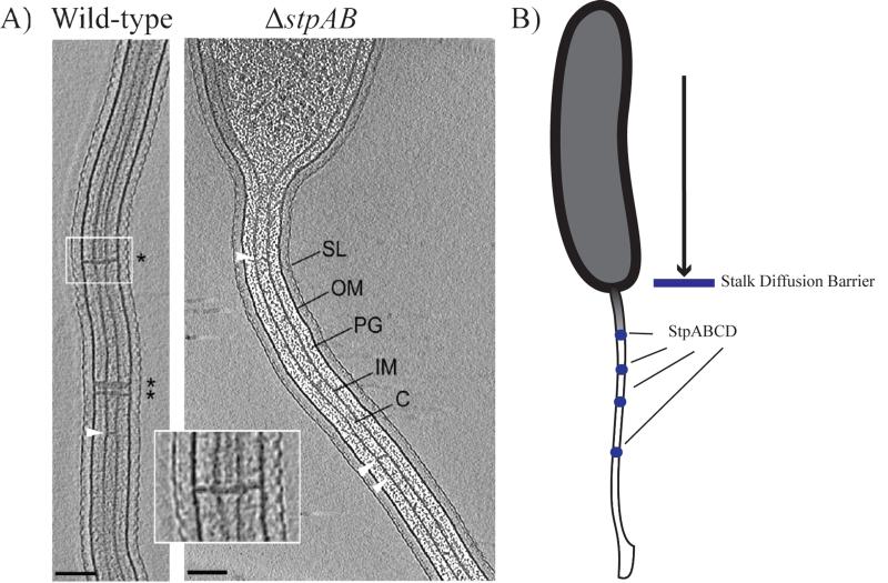
Figure 2.
A) Electron cryotomography images of wild-type Caulobacter crescentus cells containing stalk diffusion barriers (* and inset). ΔstpAB cells lack these structures. White arrows indicate unidentified structures that span the interior of the stalk (SL= S layer, OM= outer membrane, PG= peptidoglycan, IM= inner membrane, C= cytoplasm). Scale bar 100 nm. Reprinted from [Schlimpert et al., Cell, 2011] with permission from Elsevier. B) Stalk diffusion barriers: Comprising a protein complex of StpA, StpB, StpC and StpD (blue circles), diffusion barriers limit soluble and membrane protein diffusion into the stalk of C. crescentus.

