
Figure 6. Schematic diagram of sheet opening in pleurotolysin pore formation.
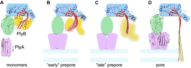
(A) PlyA and B monomer structures with coloured shapes indicating the major elements used in flexible fitting. (B) and (C) show the two types of prepore structures found, which can be considered as early and later stages of sheet opening. The top 20 models (pink for the MACPF β-sheet and grey for HTH region) and best scoring model (red) in the pleurotolysin prepore maps are shown. (B) TMH2 helix lock, in which the best scoring model has a rotation angle (β-sheet opening) of 37° relative to the monomer structure. (C) TMH2 strand lock, in which the best scoring model has a rotation angle of 49° relative to the monomer. (D) Pore model with 70° open sheet and membrane inserted TMH regions.