
Figure 4.
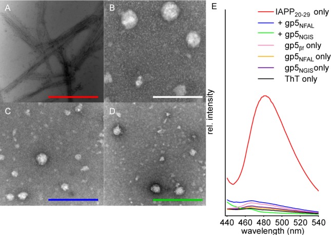
Morphological characterization of gp5βf-affected assembly of IAPP20–29. Negative stain TEM of fibers formed by 750 μM IAPP20–29 alone (A). (B–D) The same reaction as in panel A, but with the addition of 10 μM gp5βf (B), gp5NGIS (C), or gp5NFAL (D). Reaction mixtures were incubated for ∼10 h before being analyzed. Scale bars are 200 nm. (E) Fluorescence emmission spectra of 10 μM ThT added to end-state IAPP20–29 reactions of IAPP20–29 alone (red) or in the presence of gp5NFAL (blue) and gp5NGIS (green). Data for ThT alone (black) or ThT added to buffer containing only 10 μM gp5βf (pink), gp5NFAL (orange), or gp5NGIS (purple) are also shown.