
Figure 2.
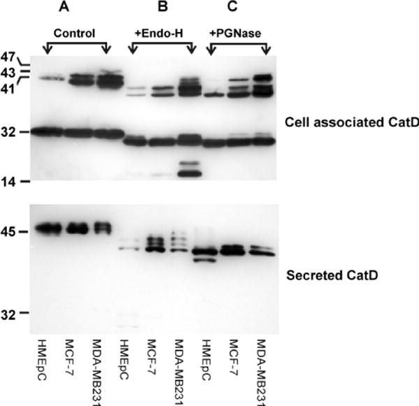
CatD cleavage products and N-glycan structures in normal mammary epithelial cells (HMEpCs) compared to breast cancer cell lines MCF-7 and MDA-MB231. Cytosolic fractions (25 μg total protein), and conditioned media (CM) from HMEpCs and breast cancer cell lines MCF-7 and MDA-MB231 were treated with or without endoglycosidase H (Endo-H, to remove the chitobiose core of high mannose and some hybrid oligosaccharides), and peptide-N-glycosidase F (PGNase, to remove high mannose, hybrid and complex glycans), then subjected to SDS-PAGE (10% acrylamide gel) and Western blot analysis. CM from the three cell lines was concentrated prior to treatment (HMEpC: 35×, MCF-7 and MDA-MB231: 2×). Differences in the abundance of the processed forms (A), and the N-glycan moieties of CatD (B&C), between normal mammary epithelial cells and breast cancer cell lines are evident in these Western blots. Note the preferential presence of multiple high mannose N-glycan structures (indicated by the appearance of multiple bands following Endo-H treatment) of the CM from the cancer cell lines.