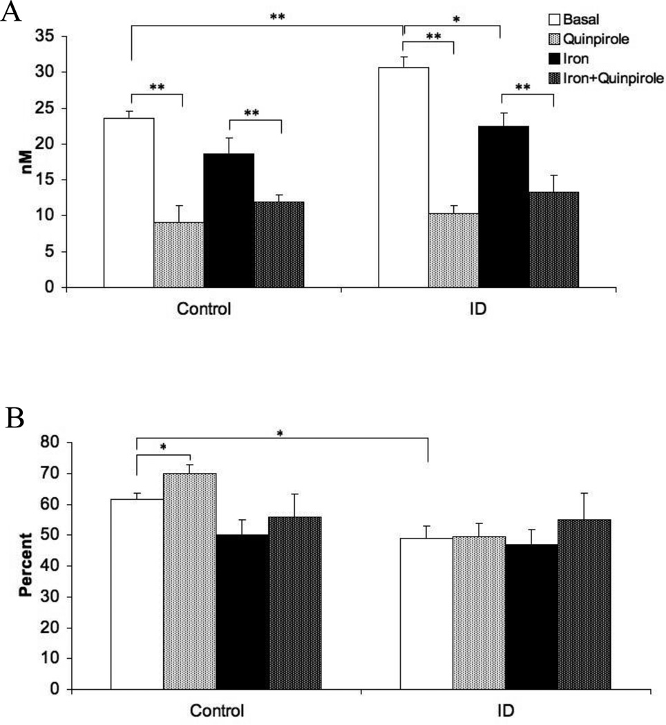
Figure 1.
(A) Extracellular dopamine concentration in the striatum before and after quinpirole perfusion into the striatum and before and after iron infusion into the midbrain. (B) Extraction fraction of extracellular DA in the striatum before and after quinpirole perfusion into the striatum and before and after iron infusion into the midbrain. Measurements were collected by in vivo microdialysis in control and iron deficient (ID) rats. Data are presented as means ± sem of 6-7 rats per group. Significance is denoted as *p < 0.05, **p < 0.01

