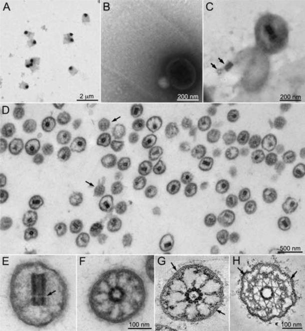
Figure 2.
Electron microscopy of isolated TZs. A. Negative stain electron microscopy of a band from a cesium chloride gradient of isolated cell walls reveals flagellar collars containing an electron dense vesicle. B. At higher magnification fibers of the flagellar collar can be seen entrapping the vesicle. C. After detergent treatment of the grid, the central cylinder characteristic of the TZ is seen inside the vesicles. The lower vesicle has lysed releasing the central cylinder, which separated into its proximal and distal components (arrows). D. Purified TZs, identified by the presence of the central cylinder in medial sections, are seen by electron microscopy of a section through a pellet of material fractionated on an iodixanol gradient. Tangential sections of TZs reveal a crosshatching of electron dense material on the inner surface of the membrane (arrows). E. In longitudinal section the distal and proximal sections of the central cylinder are visible although the transverse plate is missing (arrow). F. In cross section, although the microtubules had depolymerized, attachments can be seen between the central cylinder and the membrane. G and H: Virtual 6 nm sections of tomographic reconstructions of a TZ after isolation (G) or in situ (H). Y-shaped connectors can be seen between the outer doublet microtubules and the membrane in the TZs in situ (H, arrows). The Y-shape can sometimes be seen in the membrane attachments in the isolated TZs (G, arrows). See also Figs. S1 and S2 and Movie S1.

