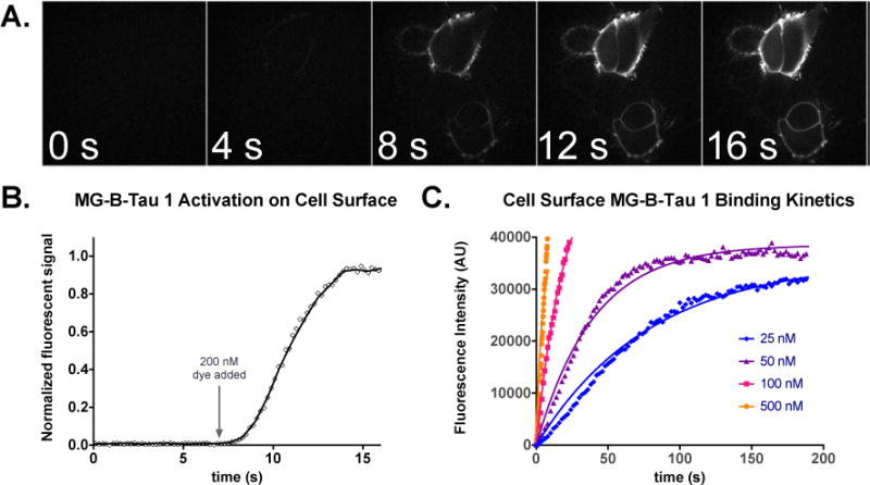
Figure 2.

Rapid activation of MG-B-Tau 1 on HEK293 cells expressing B2AR-dL5**. A. Cells were incubated in 150uL colorless DMEM and imaging was performed upon addition of 2mL of 200nM MG-B-Tau 1 at t=7 seconds to the dish until steady-state labeling was obtained. (scale bar 10 μm). B. Timecourse of activation upon addition of dye in A. Once mixed, the dye rapidly activates surface exposed FAP. C. Kinetic analysis of binding to cells at different dye concentrations and fitting to a global exponential association model reveals an activation constant of 5.06×10⁵ M−1s−1.
