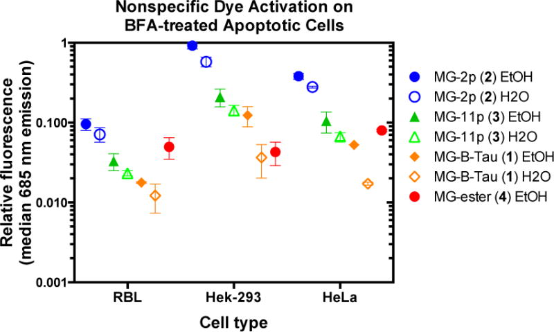
Figure 3.

Non-specific dye activation on apoptotic cells. Cells that were not expressing any FAP were treated with Brefeldin A to induce apoptosis followed by 30 minute incubation with dyes at 500 nM (MG-2p 2, MG-11p 3, and MG-B-Tau 1) or 100 nM (MG-ester 4) prepared from the indicated stock solution. Propidium iodide positive cells were selected and analyzed for associated MG fluorescence due to nonspecific activation (633 nm laser excitation with 685/70 nm emission filter). Data was normalized to the highest signal among the samples, and plotted as mean (center line) and range (box) of independent duplicate experiments on separate days.
