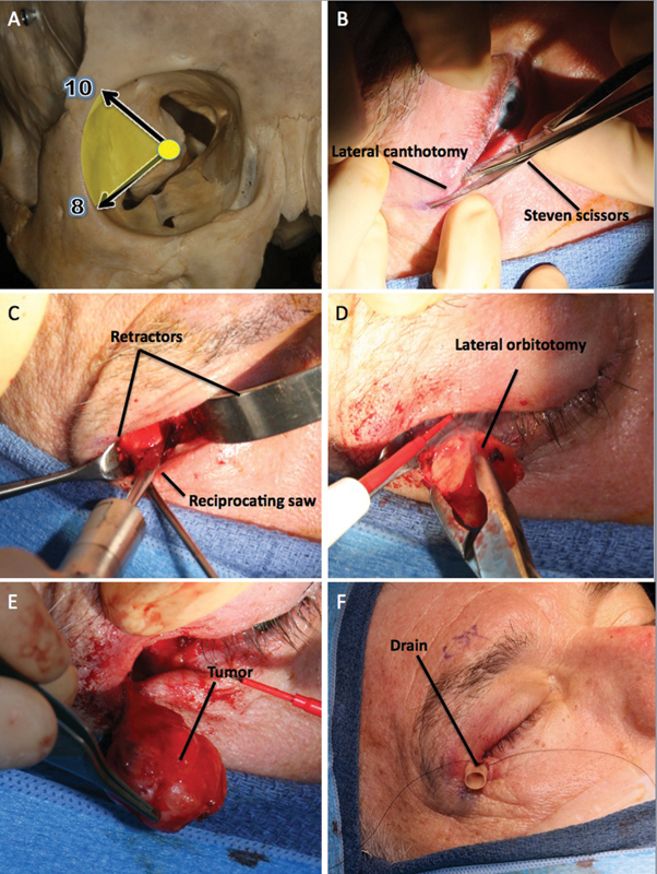
Fig. 2.

Lateral orbitotomy. (A) This approach is ideally suited for a lesion lateral to the optic nerve at the 8–10 o'clock position. (B) A small cantholysis incision is made with Steven scissors along a skin crease. (C) The temporalis muscle has been detached and retracted laterally; with adequate retraction it possible to expose the whole lateral wall of the orbit even with a relatively small incision. The osteotomy is performed with a reciprocating saw. (D) The lateral orbitotomy has been completed and the last fibers of the temporalis muscle cut with a monopolar knife. (E) The periorbita has been opened and the tumor completely removed. (F) A soft drain has been secured in place.
